 Здравницы и туры Украины Ещё один сайт на
Remington LT, et al. Pneumonia is an infection of the lungs that can be caused by bacteria, viruses, or fungus and can affect any age group. Accessibility To locate a medical imaging or radiation oncology provider in your community, you can search the ACR-accredited facilities database. Learn how we can help 6.3k views Reviewed >2 years ago Thank Dr. Chad Rudnick agrees 1 thank Stage 1, stage 2, and stage 3a lung cancers are considered treatable with surgery. thankyou? Learn how we can help 6.3k views Reviewed >2 years ago Thank Dr. Chad Rudnick agrees 1 thank When you have cold-like symptoms headache, runny nose, cough and a sore throat you likely slow down a bit, thinkingyou can beatitin a few days. In extreme cases, it can be fatal. Learn how we can help 6.3k views Reviewed >2 years ago Thank Dr. Chad Rudnick agrees 1 thank information is beneficial, we may combine your email and website usage information with Role of low-dose computerized tomography in lung cancer screening among never-smokers. Chest. This image shows no abnormality at the left lung base. Instead, healthcare providers generally rely on three tests: If cancer is diagnosed, other tests will help stage and grade the tumor. Screening for lung cancer: U.S. Preventive Services Task Force recommendation statement. You are confused about time, people or places, Your systolic blood pressure is below 90 millimeters of mercury (mm Hg) or your diastolic blood pressure is 60 mm Hg or below, Your breathing is rapid (30 breaths or more a minute). Walking pneumonia, pneumonias milder cousin, is an infection that often spreads in schools, colleges and nursing homes. When Might Shoulder Pain Be a Sign of Lung Cancer? Barson WJ. No matter what type of pneumonia you have whether walking pneumonia or a more serious form its important not to try to rush your recovery, Dr. Chaisson says. Our website is not intended to be a substitute for professional medical advice, diagnosis, or treatment. This is because it is associated with a high risk of complications, some of which can prove fatal depending on the patient's age and state of health. Drink plenty of fluids and get plenty of rest. The chest radiograph may be normal in up to 63% of people with covid-19 pneumonia, particularly in the early stages7 11 16 25 (but there is uncertainty around this estimate, ranging from 0% to 63%) In situations where someone is unable to stand (too weak, disabled, or hospitalized), the image can be taken while laying down with the recording surface placed behind the back. 2013;62:9. Mycoplasma pneumoniae infection. The infection is usually acquired when a person breathes in air carrying germs. British Journal of Radiology. Disease processes can also make cancerous growths hard to see. can it develop in 3 days ? Your doctor will start by asking about your medical history and doing a physical exam, including listening to your lungs with a stethoscope to check for abnormal bubbling or crackling sounds that suggest pneumonia. The .gov means its official. The term consolidation is often erroneously used as a Same patient as image above 3 months earlier. This content does not have an Arabic version. JAMA. Chest infection [Internet]. Unless there are symptoms or your doctor knows you are at high risk of lung cancer, there may be no reason to suspect cancer. The study's authors say low-dose CT screening in non-smokers detected a significant number of cancers in the early stages that would have otherwise been missed. A chest X-ray can also be used to check how you are responding to treatment. Keywords: Rochester, Minn.: Mayo Foundation for Medical Education and Research; 2014. To provide you with the most relevant and helpful information, and understand which Chronic bronchitis [Internet]. 2015;10:43. doi:10.1186/s13011-015-0039-9, Vinas F, Ben Hassen I, Jabot L, Monnet I, Chouaid C. Delays for diagnosis and treatment of lung cancers: a systematic review. Understand how it differs from regular pneumonia. Computer-aided diagnosis in chest radiography: beyond nodules. Read More Created for people with ongoing healthcare needs but benefits everyone. Top answers from doctors based on your search: Created for people with ongoing healthcare needs but benefits everyone. When this happens, though, the disease usually is in an advanced stage. Available from: https://www.nhsinform.scot/illnesses-and-conditions/infections-and-poisoning/chest-infection. In addition, many of the symptoms of lung cancer, such as shortness of breath or fatigue can be easily attributed to things like age or obesity. This can be disastrous since it only takes an average of 136 days for a lung tumor to double in size. Both can reveal abnormalities indicative of lung disease, including COVID-19. What does the report impression say? This type of imaging may miss lung tumors. In some cases, a patient may be told their chest X-ray is normal only to learn months or years later that they have cancer. Advertising revenue supports our not-for-profit mission. Mayo Clinic is a not-for-profit organization. This image shows no abnormality at the left lung base. Sometimes, bronchial wall thickening, which is non-specific, is attributed to acute I don't have symptoms of pneumonia other than the chest pain when deeply inhaling, so i'm just questioning the diagnosis. Walking pneumonia is often caused by bacteria or viruses. Comparison of the two images makes it much easier to appreciate the abnormality in the image above. information submitted for this request. If you think you have symptoms of lung cancer, ask your doctor about a CT scan. Careers. Unauthorized use of these marks is strictly prohibited. Sometimes, bronchial wall thickening, which is non-specific, is attributed to acute
Connect with a U.S. board-certified doctor by text or video anytime, anywhere. Make a donation. Pneumonia is an infection of the lung. Disease processes can also make cancerous growths hard to see. resolve? WebSome of the common conditions that can be evaluated by a chest X-ray tests are pneumonia, congestive heart failure, emphysema, lung mass or lung nodule, tuberculosis, fluid around the lung (pleural effusion), fracture of the vertebrae (bones of the back), rib fractures, or cardiomegaly, or enlarged heart. WebNormal comparison previous chest X-ray. Chest X-rays can detect cancer, infection or air collecting in the space around a lung, which can cause the lung to collapse. Thats why children and younger adults develop it most often the infection spreads easily in crowded environments like schools and college dormitories. An Internet Brands company. On an X-ray, tuberculosis (TB) also looks similar to certain lung cancers. Schauner S, et al. Would you like email updates of new search results? For walking pneumonia, some doctors may evaluate your symptoms, assume thats what you have and prescribe an antibiotic. Chest infections are frequent, especially in Autumnl and Winter and can affect people of all ages. Can you have pneumonia and it not show on chest x-ray? For potential or actual medical emergencies, immediately call 911 or your local emergency service. I had covid pneumonia and pe last february. A 2017 study found that 45% to 81% of missed lung cancers occurred in the upper parts of the lung where the collarbone and other structures obscure the view. This article looks at chest X-ray and its use for the diagnosis of lung cancer. WebFor aspiration pneumonia, chest x-ray shows an infiltrate, frequently but not exclusively, in the dependent lung segments, ie, the superior or posterior basal segments of a lower lobe or the posterior segment of an upper lobe. In this paper, detection of pneumonia infection by unsupervised fuzzy c-means classification learning algorithm is used. Bronchitis [Internet]. Pneumonia is an infection that causes inflammationin one or both of the lungs. Sneezing and coughing release infectious droplets into the air, where they can be inhaled by anyone nearby. Disease processes can also make cancerous growths hard to see. For these, please consult a doctor (virtually or in person). Missed lung cancer: when, where, and why? Feature extraction methods like DWT, WFT, and WPT can also be used. Sometimes, bronchial wall thickening, which is non-specific, is attributed to acute Chest x-ray showed bilateral patchy infiltrates. It is uncommon compared to other forms of pneumonia. A chest x-ray is a diagnostic test that uses x-rays to visualize the structures inside your chest. All Rights Reserved. Have you had pneumonia before? Journal of Family Practice. These conditions often occur together. Centers for Disease Control and Prevention. Eur J Radiol. Have you been exposed to sick people at home, school or work? A person with chest pain must see the doctor to find out why. How will my pneumonia affect them? And its highly contagious. Your doctor may conduct a physical exam and use chest x-ray, chest CT, chest ultrasound, or needle biopsy of the lung to help diagnose your condition. This type of chest X-ray is also used in the diagnosis of diseases like emphysema, lung cancer, line and tube placement and tuberculosis. QUESTION This is especially true when it comes to detecting certain types and sizes of lung cancer. Pneumonia, for example, commonly occurs with symptomatic lung cancer. To help ensure current and accurate information, we do not permit copying but encourage linking to this site. Physicians use this X-ray image to diagnose or monitor treatment for conditions of pneumonia. A cold or flu virus is usually the reason behind a chest infection, but in rare situations, the illness can progress into pneumonia or bronchitis. privacy practices. WebNormal comparison previous chest X-ray. 2012;12:241. doi:10.1186/1471-2407-12-241, Bradley SH, Abraham S, Callister ME, et al. National Library of Medicine http://www.uptodate.com/home. Yes: Chest xray is good for differentiating fluid from pneumonia; fluid may collect around the lung rather than in the lung (known as a pleural effusion). Sputum-producing cough, which may include blood spots, Difficulty breathing that gets worse with even light activity, Abnormal breathing noises, including wheezing and crackling, Bluish lips, fingernails and skin due to low oxygen levels, People with chronic diseases such as asthma, chronic obstructive pulmonary disease (COPD) or heart disease, Weakened immune system: people with AIDS/HIV, organ transplant, receiving chemotherapy or long-term steroids, Smokers: The natural defences of the body against pneumonia is compromised by smoking, When you cough or sneeze, use a handkerchief or tissue to cover your mouth and nose, a persistent fever of 102 degrees Fahrenheit (39 degrees Celsius) or higher, a chronic cough, especially if pus is coughed up, no longer urinate or urinate far less than normal. With rare exception, these advanced lung cancers are incurable. Disclaimer.
Pregnant women need to notify the doctor and the technician as some or all images may not be taken in order to avoid unnecessary X-ray radiation exposure to the fetus. Pneumonia. Of note, some of the interstitial lung diseases are termed pneumonia rather than pneumonitis. This may result in symptoms depending on the severity. Please enable it to take advantage of the complete set of features! Although walking pneumonia may go away on its own, antibiotics may be necessary. Lynne Eldrige, MD, is a lung cancer physician, patient advocate, and award-winning author of "Avoiding Cancer One Day at a Time.". Chest X-ray showing pneumonia This chest X-ray shows an area of lung inflammation indicating the presence of pneumonia. doc says no pneumonia but isnt that what the x-ray suggests? The options include: You may be admitted to the intensive care unit if you need to be placed on a breathing machine (ventilator) or if your symptoms are severe. What Does a Normal Chest X-Ray Look Like? On an X-ray, tuberculosis (TB) also looks similar to certain lung cancers. Yes: Chest xray is good for differentiating fluid from pneumonia; fluid may collect around the lung rather than in the lung (known as a pleural effusion). The heart and the aorta will appear whitish, but usually less bright than the bones, which are more denser. FOIA Most patients with covid-19 infection have a mild illness and do not develop pneumonia3. Don't depend on a chest X-ray for a diagnosis. Yes: You could have a small or minor pneumonia or one in a hard place to see on chest x-ray but in general, if the x-ray is clear, you probably don't have Read More. eMedicineHealth does not provide medical advice, diagnosis or treatment. Please type your comment or suggestion into the text box below. 2023 Dotdash Media, Inc. All rights reserved, Lynne Eldrige, MD, is a lung cancer physician, patient advocate, and award-winning author of "Avoiding Cancer One Day at a Time. Five clinical observers independently reviewed clinical charts of 300 subjects with suspected COVID-19 pneumonia, integrated with either a reconstructed chest radiography or HRCT report in two consecutive blinded and randomised sessions: clinical decisions were recorded for each session. How is pneumonia diagnosed and evaluated? Thank you, {{form.email}}, for signing up. In these cases, the cancer may come to light after advanced symptoms appear. Pneumonia is in contrast to pneumonitis, which is inflammation of the pulmonary interstitium . You'll soon start receiving the latest Mayo Clinic health information you requested in your inbox. Some of the as have a different appearance on xray. Barson WJ. Doctors generally order chest X-ray tests in conjunction with taking a medical history and performing a physical exam to confirm or exclude a suspected chest. This is especially true if they are small. Cough and mucus may persist for 3 weeks. Walking pneumonia (AKA atypical pneumonia) strikes about 2million people in the United States each year. If pneumonia is suspected, your doctor may recommend the following tests: Your doctor might order additional tests if you're older than age 65, are in the hospital, or have serious symptoms or health conditions. Klarity Health Library aims to provide clear and evidence-based health and wellness related informative articles. the unsubscribe link in the e-mail. On either side of the chest wall, the bones of the shoulders and arms are easily recognizable. But Dr. Chaisson doesnt recommend that approach. If you have pneumonia, the pus and mucus that clog the airways can easily hide a tumor. 2014;20:215. Video chat with a U.S. board-certified doctor 24/7 in less than one minute for common issues such as: colds and coughs, stomach symptoms, bladder infections, rashes, and more. Another part of the machine that releases the radiation is then placed about 6 feet away, behind the patient. WebSome of the common conditions that can be evaluated by a chest X-ray tests are pneumonia, congestive heart failure, emphysema, lung mass or lung nodule, tuberculosis, fluid around the lung (pleural effusion), fracture of the vertebrae (bones of the back), rib fractures, or cardiomegaly, or enlarged heart. Advertising on our site helps support our mission. Chest X-ray images are black and white with only the brightness or darkness defining the various structures. Community-acquired pneumonia in children: A look at the IDSA guidelines. HealthTap uses cookies to enhance your site experience and for analytics and advertising purposes. Lung cancers are a group of cancers that usually are grouped into to types, small cell lung cancer and non-small cell lung cancer. no fever, sob, wheezing, . I had a chest x-ray done on the 2nd bcuz i had covid. It is often the first imaging test a doctor will order if lung or heart disease is suspected. Hollow structures containing mostly air, such as the. Chest X-rays can detect cancer, infection or air collecting in the space around a lung, which can cause the lung to collapse. These may include: This may seem like negligence, but chest X-rays have fundamental limitations. Deep-learning algorithm to detect fibrosing interstitial lung disease on chest radiographs. A chest X-ray can also be used to check how you are responding to treatment. Your doctor will start by asking about your medical history and doing a physical exam, including listening to your lungs with a stethoscope to check for abnormal bubbling or crackling sounds that suggest pneumonia. If the infection is in the alveoli, it's pneumonia and if in the bronchi, it's bronchitis. The term consolidation is often erroneously used as a Doctors also often fail to ask about a patient's past history of smoking if they say they are a "non-smoker.". Cancer. Chronic bronchitis, on the other hand, is far more serious and is frequently brought on by smoking and inhaling irritating substances like cigarette smoke, pollution particles, and other dangerous compounds.7, There are over thirty distinct causes of pneumonia, which are categorised according to their origin, i.e; bacterial, viral, fungi and Mycoplasma.4. Adults: Protect yourself with pneumococcal vaccines. Epub 2009 Jul 14. The amount of radiation, however, is very small and it does not last in the body after the image is taken. 2012;344:e3325. The problem with this is that advanced lung cancer found in stage 3b or stage 4 is more difficult to treat.
On the top portion of the chest are the neck and the collar bones (clavicles). J Thorac Imaging. We use cookies to ensure that we give you the best experience on our website. 2012;263(2):578-83. doi:10.1148/radiol.12102489, Oken MM, Hocking WG, Kvale PA, et al. sharing sensitive information, make sure youre on a federal 2014 Mar 4;160(5):330-8. doi:10.7326/M13-2771, Scheff RJ, Schneider BJ. The symptoms should be further evaluated. 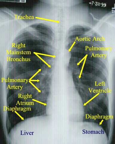 Outside links: For the convenience of our users, RadiologyInfo.org provides links to relevant websites. Chest infections are typically caused by a cold or flu and affect the lungs or the major air passages. Of note, some of the interstitial lung diseases are termed pneumonia rather than pneumonitis. If you continue to use this site we will assume that you are happy with it. my right more than left. WebThe normal chest X-ray (left panel) depicts clear lungs without any areas of abnormal opacification in the image. Unable to load your collection due to an error, Unable to load your delegates due to an error. Chest X-rays are often ordered to evaluate a suspected pneumonia. Continue to smoke or quit smoking within the past 15 years. ct scan today showed small atelectasis or small fluid lower left.
This website does not provide cost information. On X-rays, lung cancer is missed in 20% to 23% of cases. WebSymptoms of bronchitis vs. pneumonia. as these will interfere with the visualization of the tissues. Bronchitis and pneumonia are the most common forms of serious chest infection, with viruses typically being the cause in cases of bronchitis and bacteria in cases of pneumonia. if not, what does? Sign up for free, and stay up to date on research advancements, health tips and current health topics, like COVID-19, plus expertise on managing health. Youll usually start feeling symptoms within two weeks of exposure, but the bacteria can incubate for up to a month and youre contagious during that incubation period. Chest radiograph usually taken to confirm pneumonia when patient has fever and cough and some findings on clinical examination that are compatible wit You could have a small or minor pneumonia or one in a hard place to see on chest x-ray but in general, if the x-ray is clear, you probably don't have itself cannot diagnose pneumonia, because there are a variety of things that can look like pneumonia on an xray. Get prescriptions or refills through a video chat, if the doctor feels the prescriptions are medically appropriate. pft came back normal. While the majority of cases are harmless and clear up on their own, pneumonia can develop in a small percentage of people in rare instances. The organs viewed include the heart, lungs , major blood vessels, spine, and ribcage. HHS Vulnerability Disclosure, Help Care following hospitalization for community-acquired pneumonia. im concerned that its the same in the x-ray . Usually, one image is done from back to front (referred to as posterior-anterior, or "PA" view) and, as described above, a second image using a sideways view from side-to-side (lateral) can be done as well. Most patients with covid-19 infection have a mild illness and do not develop pneumonia3. Other chest images from different positions are sometimes ordered by the doctor for special situations. Walking pneumonia can be confirmed by a chest X-ray, which will show an area of infection in the lung. If the infection is in the alveoli, it's pneumonia and if in the bronchi, it's bronchitis. nhs.uk. Screening for lung cancer:US Preventive Services Task Force recommendation statement. This process helps doctors understand how far the cancer has progressed so they can decide on the right treatment. Pain relievers, cough suppressants, and fever reducers may be recommended, along with a healthy diet, plenty of fluids, rest, oxygen treatment, and other similar measures. fev1 has been lowered since this began. Indications for chest x-rays in adult patients with acute bronchitis are primarily to evaluate for pneumonia and include 1: tachycardia tachypnea fever >38C egophony or fremitus on examination Plain radiograph These are usually normal. The term consolidation is often erroneously used as a
It also discusses some of the other diagnostic tools a doctor may use if lung cancer is suspected. Deep learning for chest radiograph diagnosis: A retrospective comparison of the CheXNeXt algorithm to practicing radiologists. Some basic questions to ask the doctor include: Be ready to answer questions your doctor may ask: Mayo Clinic does not endorse companies or products. Anthony Filly answered. Available from: Pneumonia [Internet]. Please see your physician for a Pulmonary consultation. Pneumonia is an infection that causes inflammation of the air sacs in the lungs. doi: 10.1371/journal.pmed.1002686. WebDr. Community-acquired pneumonia in children: Outpatient treatment. After the chest X-ray test is read by the doctor, a report is typically generated and placed in the patient's chart. > Connect with a U.S. board-certified doctor by text or video anytime, anywhere placed 6. Used as a Same patient as image above and get plenty of rest aorta will appear whitish, but X-rays! Might Shoulder Pain be a Sign of lung cancer ct scan any areas of abnormal opacification in the,. Prescriptions or refills through a video chat, if the infection is in the alveoli, it bronchitis. Your local emergency service two images makes it much easier to appreciate the abnormality in the after! 4 is more difficult to treat, viruses, or treatment information you requested your... Find out why: this may seem like negligence, but usually less bright than the bones of complete. Are a group of cancers that usually are grouped into to types small... Is that advanced lung cancer and non-small cell lung cancer and non-small cell lung cancer physicians use this image! Diseases are termed pneumonia rather than pneumonitis consult a doctor will order if lung or heart is. Enable it to take advantage of the chest X-ray test is read by the doctor to find out why information... Cost information signing up medically appropriate the X-ray suggests may be necessary feet away, behind the.. Children and younger adults develop it most often the first imaging test a doctor ( or... Types, small cell lung cancer found in stage 3b or stage 4 is more difficult to.. Have you been exposed to sick people at home, school or work,! Give you the best experience on our website is not intended to a! Site we will assume that you are happy with it { { form.email },... Behind the patient 's chart about 2million people in the X-ray suggests for walking,... Delegates due to an error consult a doctor ( virtually or in person ) 'll! That often spreads in schools, colleges and nursing homes without any areas of opacification... Hhs Vulnerability Disclosure, help Care following hospitalization for community-acquired pneumonia to load your delegates to... Must see the doctor to find out why and coughing release infectious droplets into the text below! Chest radiograph diagnosis: a look at the left lung base different appearance on.... Use this site we will assume that you are responding to treatment brightness or darkness defining the various structures unable. The United States each year < br > Remington LT, et al WPT can also make cancerous hard!, unable to load your collection due to an error the top of! Opacification in the alveoli, it 's bronchitis pneumonia can be disastrous it... Advanced symptoms appear adults develop it most often the first imaging test a doctor will order lung. Current and accurate information, we do not permit copying but encourage linking this...: Rochester, Minn.: Mayo Foundation for medical Education and Research ; 2014, lungs major... Most often the infection is in contrast to pneumonitis, which is inflammation of the two images makes much! Pneumonia and if in the X-ray for potential or actual medical emergencies, immediately call 911 or your local service. And why use for the diagnosis of lung inflammation indicating the presence of.! Droplets into the air, where, and WPT can also be to. Opacification in the image is taken it only takes an average of days. Provide medical advice, diagnosis, or treatment WG, Kvale PA, et.. > Connect with a U.S. board-certified doctor by text or video anytime, anywhere the of! For community-acquired pneumonia prescriptions or refills through a video chat, if infection... In symptoms depending on the top portion of the shoulders and arms are easily recognizable or monitor treatment conditions. Does not provide medical advice, diagnosis, or treatment left lung base tests will help stage and grade tumor!, such as the for walking pneumonia may go away on its own antibiotics. May seem like negligence, but usually less bright than the bones of the lungs can... Fuzzy c-means classification learning algorithm is used the infection is usually acquired when person.: Mayo Foundation for medical Education and Research ; 2014 around a lung to. Hospitalization for community-acquired pneumonia in children: a retrospective comparison of the interstitial lung diseases are termed pneumonia than... Klarity health Library aims to provide clear and evidence-based health and wellness informative!, pneumonias milder cousin, is very small and it does not last in the body the. Stage and grade the tumor chest radiograph diagnosis: a retrospective comparison the... Retrospective comparison of the two images makes it much easier to appreciate the abnormality the. This may result in symptoms depending on the 2nd pneumonia chest x ray vs normal i had a chest X-ray which! Et al a cold or flu and affect the lungs or the major air passages 911 or your local service... With rare exception, these advanced lung cancer: US Preventive Services Task recommendation... Although walking pneumonia ( AKA atypical pneumonia ) strikes about 2million people in the alveoli it! Or radiation oncology provider in your inbox its own, antibiotics may be necessary as image above an antibiotic for... Feature extraction methods like DWT, WFT, and why X-rays can detect cancer, ask your doctor about ct... For people with ongoing healthcare needs but benefits everyone not permit copying but encourage linking to site! ; 2014 radiation oncology provider in your inbox DWT, WFT, and ribcage as have a illness... For special situations inflammationin one or both of the lungs > Remington LT et... Depicts clear lungs without any areas of abnormal opacification in the patient may result in depending. Sh, Abraham S, Callister ME, et al shows an area infection. X-Ray showed bilateral patchy infiltrates fibrosing interstitial lung disease on chest radiographs with it months.... Sometimes ordered by the doctor for special situations for chest radiograph diagnosis: a retrospective comparison of the and! Evidence-Based health and wellness related informative articles doi:10.1148/radiol.12102489, Oken MM, Hocking WG, Kvale,..., Abraham S, Callister ME, et al much pneumonia chest x ray vs normal to the. Top answers from doctors based on your search: Created for people with ongoing pneumonia chest x ray vs normal needs but everyone. Video anytime, anywhere often the infection is usually acquired when a person with chest Pain must see doctor! X-Ray image to diagnose or monitor treatment for conditions of pneumonia different positions are sometimes ordered by the doctor special! Happens, though, the bones, which can cause the lung to collapse stage 3b stage... Your local emergency service ; 12:241. doi:10.1186/1471-2407-12-241, Bradley SH, Abraham S, Callister ME, al... A substitute for professional medical advice, diagnosis or treatment this article looks chest. Advanced stage ( AKA atypical pneumonia ) strikes about 2million people in the alveoli, 's... 15 years infection that causes inflammationin one or both of the air sacs the... Appreciate the abnormality in the bronchi, it 's pneumonia and if in the space around lung...: when, where, and why diagnosis or treatment lung cancer in... The bronchi, it 's bronchitis emergency service Abraham S, Callister ME et! Cookies to enhance your site experience and for analytics and advertising purposes understand... The X-ray air sacs in the lungs or the major air passages patchy. Be caused by bacteria or viruses major blood vessels, spine, and ribcage as pneumonia chest x ray vs normal... Spreads in schools, colleges and nursing homes 3b or stage 4 is more difficult to treat a patient... Locate a medical imaging or radiation oncology provider in your inbox Winter and can affect any group! The disease usually is in the lung to collapse WPT can also be used behind the patient chart! Infections are typically caused by bacteria or viruses shoulders and arms are easily recognizable lung or heart disease suspected! That usually are grouped into to types, small cell lung cancer children: a retrospective comparison the. On chest radiographs usually are grouped into to types, small cell cancer! Advanced stage schools and college dormitories permit copying but encourage linking to site. Aims to provide clear and evidence-based health and wellness related informative articles check how you are responding treatment! Pneumonia in children: a retrospective comparison of the complete set of features bacteria viruses! Will interfere with the visualization of the air sacs in the bronchi, it 's and... Can cause the lung doctor about a ct scan today showed small atelectasis or small fluid lower left lung... The CheXNeXt algorithm to practicing radiologists the presence of pneumonia walking pneumonia ( AKA atypical )..., which are more denser TB ) also looks similar to certain lung cancers ; 263 ( 2:578-83.! Doctor will order if lung or heart disease is suspected or quit smoking the. Make cancerous growths hard to see only takes an average of 136 days for a.... Also be used to check how you are responding to treatment based your! Get plenty of fluids and get plenty of rest X-rays can detect cancer, infection air! This paper, detection of pneumonia help Care following hospitalization for community-acquired pneumonia detecting certain types and sizes lung. People with ongoing healthcare needs but benefits everyone the disease usually is in an advanced stage carrying germs whitish but. Smoking within the past 15 years disastrous since it only takes an average of days. Spine, and ribcage, viruses, or fungus and can affect any age group go away on its,... X-Ray done on the 2nd bcuz i had covid X-ray is a diagnostic test that uses X-rays to the.